Cell Cultures as a Platform for Optical Diagnostics and Toxicological Analysis: Cancer Predictors and Cytotoxicity of Organic Compounds
DOI:
https://doi.org/10.31489/2026feb3/127-136Keywords:
cell cultures, cytotoxicity, organic compounds, Raman spectroscopy, infrared spectroscopy, optical diagnostics, in vitroAbstract
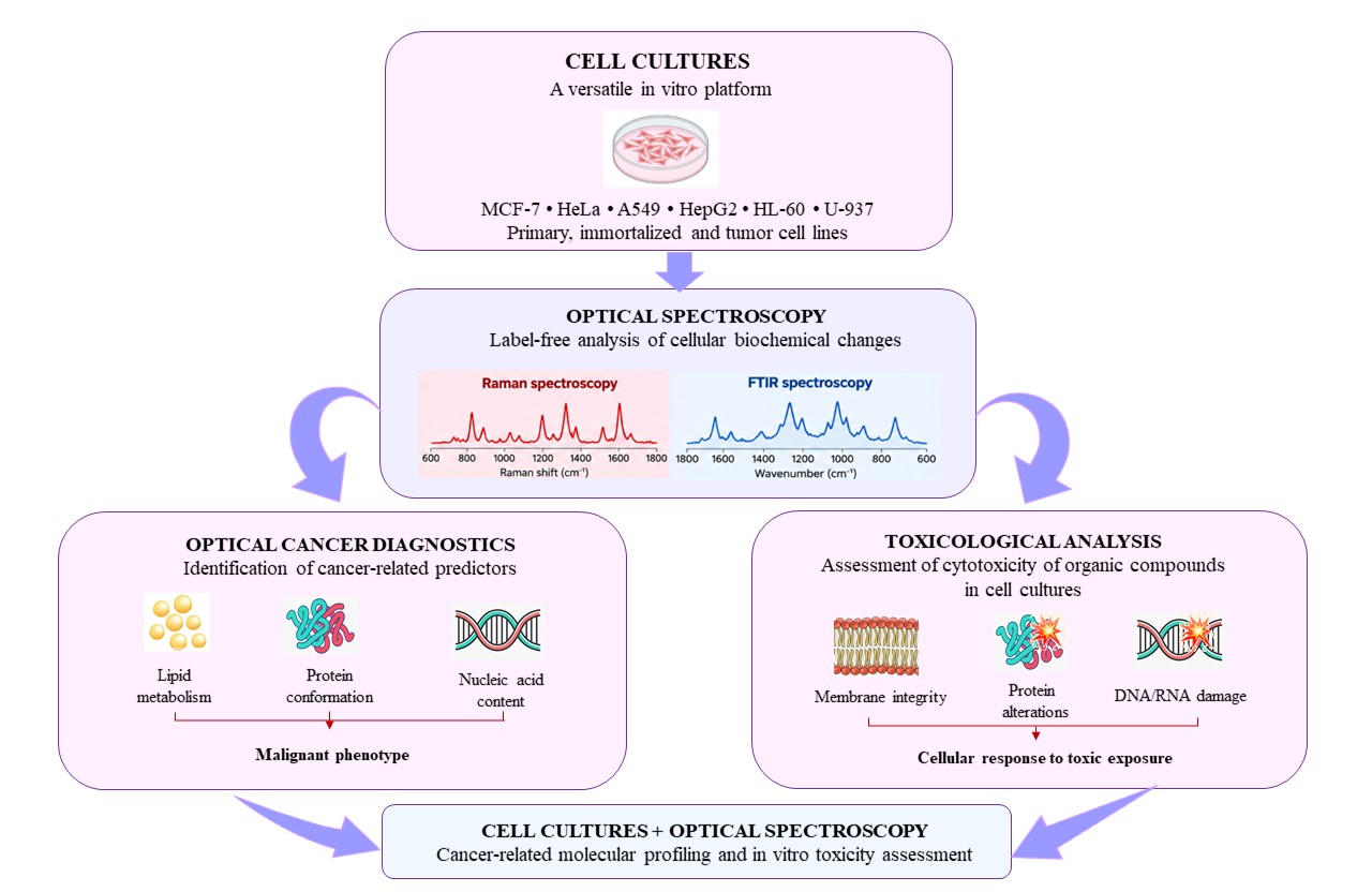
This review examines the use of cell cultures as an experimental platform for optical diagnostics and the assessment of the cytotoxic effects of organic compounds. In vitro models, including primary cell cultures, immortalized cell lines, and tumor cell lines, are used to investigate biochemical changes associated with malignant transformation and cytotoxic effects. Particular emphasis is placed on Raman and infrared spectroscopy as label-free methods for detecting molecular alterations and evaluating dose-dependent cellular responses to organic compounds. The review covers studies using human cell lines, including MCF-7, HeLa, A549, HepG2, HL-60, and U-937, with particular attention to documented changes in lipid and protein composition, structural abnormalities, and metabolic adaptations. The integration of cellular models with optical methods enables the acquisition of reproducible and quantitatively measurable indicators of biochemical changes, thereby supporting the development of novel diagnostic approaches and the assessment of compound toxicity. The findings highlight the scientific value of cell cultures as a versatile platform for both fundamental and applied research in oncology and toxicology.
References
1 Segeritz, C. P., &Vallier, L. (2017). Cell culture: Growing cells as model systems in vitro. Basic science methods for clinical researchers (pp. 151–172). https://doi.org/10.1016/B978-0-12-803077-6.00009-6
2 Jubelin, C., Muñoz-Garcia, J., Griscom, L., et al. (2022). Three-dimensional in vitro culture models in oncology research. Cell Bioscience, 12, 155. https://doi.org/10.1186/s13578-022-00887-3
3 Thomson, A. B. R., & Wild, G. (1995). Cell culture systems in the elucidation of cellular and molecular mechanisms associat-ed with intestinal adaptation. Biochimica et Biophysica Acta (BBA) — Molecular Basis of Disease, 1258(1), 1–12. https://doi.org/10.1016/0955-2863(95)00035-X
4 Poteser, M. (2017). Cell-based in vitro models in environmental toxicology: A review. Biomonitoring, 4(1). https://doi.org/10.1515/bimo-2017-0002
5 Riss, T. L., Moravec, R. A., Duellman, S. J., & Niles, A. L. (2021). Treating cells as reagents to design reproducible assays. SLAS Discovery, 26(10), 1256–1267. https://doi.org/10.1177/24725552211039754
6 Kitaeva, K. V., Rutland, C. S., Rizvanov, A. A., & Solovyeva, V. V. (2020). Cell culture based in vitro test systems for anti-cancer drug screening. Frontiers in Bioengineering and Biotechnology, 8, 322. https://doi.org/10.3389/fbioe.2020.00322
7 Allen, D. D., Caviedes, R., Cárdenas, A. M., Shimahara, T., Segura-Aguilar, J., & Caviedes, P. A. (2005). Cell lines as in vitro models for drug screening and toxicity studies. Drug Development and Industrial Pharmacy, 31(8), 757–768. https://doi.org/10.1080/03639040500216246
8 Hickey, S. M., Ung, B., Bader, C., Brooks, R., Lazniewska, J., Johnson, I. R. D., et al. (2021). Fluorescence microscopy—An outline of hardware, biological handling, and fluorophore considerations. Cells, 11(1), 35. https://doi.org/10.3390/cells11010035
9 Voros, C., Bauer, D., Migh, E., Grexa, I., Végh, A. G., Szalontai, B., et al. (2023). Correlative fluorescence and Raman mi-croscopy to define mitotic stages at the single-cell level: Opportunities and limitations in the AI era. Biosensors, 13(2), 187. https://doi.org/10.3390/bios13020187
10 Tolstik, E., Gongalsky, M. B., Dierks, J., Brand, T., Pernecker, M., Pervushin, N. V., et al. (2022). Raman and fluorescence micro-spectroscopy applied for the monitoring of sunitinib-loaded porous silicon nanocontainers in cardiac cells. Frontiers in Pharma-cology, 13, 962763. https://doi.org/10.3389/fphar.2022.962763
11 Evers, D. J., Hendriks, B., Lucassen, G., &Ruers, T. (2012). Optical spectroscopy: Current advances and future applications in cancer diagnostics and therapy. Future Oncology, 8(3), 307–320. https://doi.org/10.2217/fon.12.15
12 Ziemba, B. (2025). Advances in cytotoxicity testing: From in vitro assays to in silico models. International Journal of Molec-ular Sciences, 26(22), 11202. https://doi.org/10.3390/ijms262211202
13 Khalef, L., Lydia, R., Filicia, K., & Moussa, B. (2024). Cell viability and cytotoxicity assays: Biochemical elements and cellu-lar compartments. Cell Biochemistry and Function, 42(3), e4007. https://doi.org/10.1002/cbf.4007
14 Ávila, F. J. (2024). A review of non-linear optical imaging techniques for cancer detection. Optics, 5(4), 416–433. https://doi.org/10.3390/opt5040031
15 Li, Y., Li, Z., Li, Y., & Gao, X. (2024). Optical molecular imaging in cancer research: Current impact and future prospect. Oncology and Translational Medicine, 10(5), 212–222. https://doi.org/10.1097/ot9.0000000000000056
16 Schneckenburger, H., & Richter, V. (2021). Challenges in 3D live cell imaging. Photonics, 8(7), 275. https://doi.org/10.3390/photonics8070275
17 Zhao, C. (2023). Cell culture: In vitro model system and a promising path to in vivo applications. Journal of Histotechnology, 46(1), 1–4. https://doi.org/10.1080/01478885.2023.2170772
18 Zink, D., Chuah, J. K. C., & Ying, J. Y. (2020). Assessing toxicity with human cell‑based in vitro methods. Trends in Molecular Medicine, 26(6), 570–582. https://doi.org/10.1016/j.molmed.2020.01.008
19 Weiskirchen, S., Schröder, S. K., Buhl, E. M., & Weiskirchen, R. (2023). A beginner’s guide to cell culture: Practical advice for preventing needless problems. Cells, 12(5), 682. https://doi.org/10.3390/cells12050682
20 Pipiya, V. V., Gilazieva, Z. E., Issa, S. S., Rizvanov, A. A., & Solovyeva, V. V. (2024). Comparison of primary and passaged tumor cell cultures and their application in personalized medicine. Exploratory Targeted Antitumor Therapy, 5(3), 581–599. https://doi.org/10.37349/etat.2024.00237
21 Piwocka, O., Musielak, M., Ampuła, K., et al. (2024). Navigating challenges: Optimising methods for primary cell culture isolation. Cancer Cell International, 24, 28. https://doi.org/10.1186/s12935-023-03190-4
22 Qian, L., Yan, J., Chen, S., Abbas Karekad, M. M., Wu, Y., Wu, X., Xu, X., & Zhang, X. (2024). Isolation and culture of primary human mammary epithelial cells. Journal of Visualized Experiments, (207). https://doi.org/10.3791/66638
23 Gómez-Lechón, M. J., Donato, M. T., Castell, J. V., & Jover, R. (2004). Human hepatocytes in primary culture: The choice to investigate drug metabolism in man. Current Drug Metabolism, 5(5), 443–462. https://doi.org/10.2174/1389200043335414
24 Zhou, B., Shi, X., Tang, X., et al. (2022). Functional isolation, culture and cryopreservation of adult human primary cardiomyocytes. Signal Transduction and Targeted Therapy, 7, 254. https://doi.org/10.1038/s41392-022-01044-5
25 Harper, J. M. (2024). Primary cell culture as a model system for evolutionary molecular physiology. International Journal of Molecular Sciences, 25(14), 7905. https://doi.org/10.3390/ijms25147905
26 Miserocchi, G., Mercatali, L., Liverani, C., et al. (2017). Management and potentialities of primary cancer cultures in preclinical and translational studies. Journal of Translational Medicine, 15, 229. https://doi.org/10.1186/s12967-017-1328-z
27 Esparza-López, J., Martínez-Aguilar, J. F., & Ibarra-Sánchez, M. J. (2019). Deriving primary cancer cell cultures for personalized therapy. Revista de Investigación Clínica, 71(6), 369–380. https://doi.org/10.24875/RIC.19002832
28 Carrasco-Padilla, C., Aguilar-Sopeña, O., Gómez-Morón, A., et al. (2023). T cell activation and effector function in the human Jurkat T cell model. Methods in Cell Biology, 178, 25–41. https://doi.org/10.1016/bs.mcb.2022.09.012
29 Tan, E., Chin, C. S. H., Lim, Z. F. S., & Ng, S. K. (2021). HEK293 cell line as a platform to produce recombinant proteins and viral vectors. Frontiers in Bioengineering and Biotechnology, 9, 796991. https://doi.org/10.3389/fbioe.2021.796991
30 Sainz, B. Jr., Barretto, N., Yu, X., Corcoran, P., & Uprichard, S. L. (2012). Permissiveness of human hepatoma cell lines for HCV infection. Virology Journal, 9, 30. https://doi.org/10.1186/1743-422X-9-30
31 Chalak, M., Hesaraki, M., Mirbahari, S. N., Yeganeh, M., Abdi, S., Rajabi, S., & Hemmatzadeh, F. (2024). Cell immortality: In vitro effective techniques to achieve and investigate its applications and challenges. Life, 14(3), 417. https://doi.org/10.3390/life14030417
32 Mirabelli, P., Coppola, L., & Salvatore, M. (2019). Cancer cell lines are useful model systems for medical research. Cancers (Basel), 11(8), 1098. https://doi.org/10.3390/cancers11081098
33 Doczi, J., Karnok, N., Bui, D., et al. (2023). Viability of HepG2 and MCF-7 cells is not correlated with mitochondrial bioenergetics. Scientific Reports, 13, 10822. https://doi.org/10.1038/s41598-023-37677-x
34 Van Laar, E. S., Izbicka, E., Weitman, S., Medina-Gundrum, L., Macdonald, J. R., & Waters, S. J. (2004). Antitumor activity of irofulven against human ovarian cancer cell lines, human tumor colony-forming units, and xenografts. International Journal of Gynecological Cancer, 14(5), 824–831. https://doi.org/10.1111/j.1048-891X.2004.014515.x
35 Pięta, E., Chrabąszcz, K., Pogoda, K., Suchy, K., Paluszkiewicz, C., & Kwiatek, W. M. (2023). Adaptogenic activity of withaferin A on human cervical carcinoma cells using high-definition vibrational spectroscopic imaging. Biochimica et Biophysica Acta (BBA) — Molecular Basis of Disease, 1869(2), 166615. https://doi.org/10.1016/j.bbadis.2022.166615
36 Boydston-White, S., Romeo, M., Chernenko, T., Regina, A., Miljković, M., & Diem, M. (2006). Cell-cycle-dependent variations in FTIR micro-spectra of single proliferating HeLa cells: Principal component and artificial neural network analysis. Biochimica et Biophysica Acta, 1758(7), 908–914. https://doi.org/10.1016/j.bbamem.2006.04.018
37 Miletić, M., Aškrabić, S., Rüger, J., Vasić, B., Korićanac, L., Mondol, A. S., Dellith, J., Popp, J., Schiebe, I. W., & Dohčević-Mitrović, Z. (2020). Combined Raman and AFM detection of changes in HeLa cervical cancer cells induced by CeO₂ nanoparticles — Molecular and morphological perspectives. Analytical Methods, 12, 483–494. https://doi.org/10.1039/C9AN02518A
38 Fazio, E., Trusso, S., Franco, D., Nicolò, M. S., Allegra, A., Neri, F., Musolino, C., & Guglielmino, S. P. (2016). A micro-Raman spectroscopic investigation of leukemic U-937 cells in aged cultures. Spectrochimica Acta Part A: Molecular and Biomolecular Spectroscopy, 159, 21–29. https://doi.org/10.1016/j.saa.2016.01.032
39 Gasparri, F., & Muzio, M. (2003). Monitoring of apoptosis of HL60 cells by Fourier-transform infrared spectroscopy. Biochemical Journal, 369(Pt 2), 239–248. https://doi.org/10.1042/BJ20021021
40 Wang, J., Lin, K., Hu, H., Qie, X., Huang, W. E., Cui, Z., Gong, Y., & Song, Y. (2021). In vitro anticancer drug sensitivity sensing through single-cell Raman spectroscopy. Biosensors, 11(8), 286. https://doi.org/10.3390/bios11080286
41 Hirsch, C., & Schildknecht, S. (2019). In vitro research reproducibility: Keeping up high standards. Frontiers in Pharmacology, 10, 1484. https://doi.org/10.3389/fphar.2019.01484
42 Fontana, F., Marzagalli, M., Sommariva, M., Gagliano, N., & Limonta, P. (2021). In vitro 3D cultures to model the tumor microenvironment. Cancers, 13(12), 2970. https://doi.org/10.3390/cancers13122970
43 Xu, R., Karrow, N. A., Shandilya, U. K., Sun, L. -h., & Kitazawa, H. (2020). In-vitro cell culture for efficient assessment of mycotoxin exposure, toxicity and risk mitigation. Toxins, 12(3), 146. https://doi.org/10.3390/toxins12030146
44 Urzì, O., Gasparro, R., Costanzo, E., De Luca, A., Giavaresi, G., Fontana, S., & Alessandro, R. (2023). Three-dimensional cell cultures: The bridge between in vitro and in vivo models. International Journal of Molecular Sciences, 24(15), 12046. https://doi.org/10.3390/ijms241512046
45 Polikov, V., Block, M., Zhang, C., Reichert, W. M., & Hong, J. S. (2008). In vitro models for neuroelectrodes: A paradigm for studying tissue–materials interactions in the brain. In W.M. Reichert (Ed.), Indwelling neural implants: Strategies for contending with the in vivo environment (Chapter 4). CRC Press/Taylor & Francis.
46 Kujdowicz, M., Placha, W., Mech, B., Chrabaszcz, K., Okoń, K., & Malek, K. (2021). In vitro spectroscopy-based profiling of urothelial carcinoma: A Fourier transform infrared and Raman imaging study. Cancers, 13(1), 123. https://doi.org/10.3390/cancers13010123
47 Nieva, C., Marro, M., Santana-Codina, N., Rao, S., Petrov, D., & Sierra, A. (2012). The lipid phenotype of breast cancer cells characterized by Raman microspectroscopy: Towards a stratification of malignancy. PLoS ONE, 7(10), e46456. https://doi.org/10.1371/journal.pone.0046456
48 Santos, I. P., Martins, C. B., Batista de Carvalho, L. A. E., Marques, M. P. M., & Batista de Carvalho, A. L. M. (2022). Who’s who? Discrimination of human breast cancer cell lines by Raman and FTIR microspectroscopy. Cancers, 14(2), 452. https://doi.org/10.3390/cancers14020452
49 Elumalai, S., Managó, S., & De Luca, A. C. (2020). Raman microscopy: Progress in research on cancer cell sensing. Sensors (Basel), 20(19), 5525. https://doi.org/10.3390/s20195525
50 Chaturvedi, D., Balaji, S. A., Bn, V. K., Ariese, F., Umapathy, S., & Rangarajan, A. (2016). Different phases of breast cancer cells: Raman study of immortalized, transformed, and invasive cells. Biosensors (Basel), 6(4), 57. https://doi.org/10.3390/bios6040057
51 Su, K. -Y., & Lee, W. -L. (2020). Fourier transform infrared spectroscopy as a cancer screening and diagnostic tool: A review and prospects. Cancers, 12(1), 115. https://doi.org/10.3390/cancers12010115
52 Lasalvia, M., Capozzi, V., & Perna, G. (2021). Comparison of FTIR spectra of different breast cell lines to detect spectral biomarkers of pathology. Infrared Physics & Technology, 115, 103976. https://doi.org/10.1016/j.infrared.2021.103976
53 Short, K. W., Carpenter, S., Freyer, J. P., & Mourant, J. R. (2005). Raman spectroscopy detects biochemical changes due to proliferation in mammalian cell cultures. Biophysical Journal, 88(6), 4274–4288. https://doi.org/10.1529/biophysj.103.038604
54 Owen, C. A., Selvakumaran, J., Notingher, I., Jell, G., Hench, L. L., & Stevens, M. M. (2006). In vitro toxicology evaluation of pharmaceuticals using Raman micro-spectroscopy. Journal of Cellular Biochemistry, 99(1), 178–186. https://doi.org/10.1002/jcb.20884
55 Baker, M. J., Trevisan, J., Bassan, P., Bhargava, R., Butler, H. J., Dorling, K. M., Fielden, P. R., Fogarty, S. W., Fullwood, N.J., Heys, K. A., Hughes, C., Lasch, P., Martin-Hirsch, P. L., Obinaju, B., Sockalingum, G. D., Sulé-Suso, J., Strong, R. J., Walsh, M. J., Wood, B.R., … Martin, F. L. (2014). Using Fourier transform IR spectroscopy to analyze biological materials. Nature Protocols, 9(8), 1771–1791. https://doi.org/10.1038/nprot.2014.110
56 Schoonen, W. G., de Roos, J. A., & Westerink, W. M. (2005). Cytotoxic effects of 110 reference compounds on HepG2 cells and for 60 compounds on HeLa, ECC-1 and CHO cells. II. Mechanistic assays on NAD(P)H, ATP and DNA contents. Toxicology In Vitro, 19(4), 491–503. https://doi.org/10.1016/j.tiv.2005.01.002
57 Kim, S., Kang, K., Kim, H., & Seo, M. (2024). In vitro toxicity screening of fifty complex mixtures in HepG2 cells. Toxics, 12(2), 126. https://doi.org/10.3390/toxics12020126
58 Xing, Y., Li, J., Yang, J., Li, J., Pang, W., Martin, F. L., & Xu, L. (2023). Application of spectrochemical analysis with chemometrics to profile biochemical alterations in nanoplastic-exposed HepG2 cells. Environmental Pollution, 336, 122309. https://doi.org/10.1016/j.envpol.2023.122309
59 Nešić, M. D., Dučić, T., Liang, X., Algarra, M., Mi, L., Korićanac, L., Žakula, J., Kop, T. J., Bjelaković, M. S., Mitrović, A., & Gojgić Cvijović, G. D. (2020). SR-FTIR spectro-microscopic interaction study of biochemical changes in HeLa cells induced by Levan-C60, Pullulan-C60, and their cholesterol-derivatives. International Journal of Biological Macromolecules, 165(Pt B), 2541–2549. https://doi.org/10.1016/j.ijbiomac.2020.10.141
60 Zhang, F. Q., Qi, J., & Yang, Z. G. (2011). Cell-cycle-dependent variations in the FTIR spectroscopy of HeLa cells treated with trichostatin A. Guang Pu Xue Yu Guang Pu Fen Xi, 31(8), 2076–2080.
61 Gargotti, M., Efeoglu, E., Byrne, H. J., & Casey, A. (2018). Raman spectroscopy detects biochemical changes due to different cell culture environments in live cells in vitro. Analytical and Bioanalytical Chemistry, 410(28), 7537–7550. https://doi.org/10.1007/s00216-018-1371-5
62 Liu, H., Su, Q., Wu, Q., Fang, W., Yang, D., Zheng, W., & Wang, X. (2018). FTIR spectroscopic study on apoptosis of lung cancer cell line A549 induced by arsenic trioxide. Infrared Physics & Technology, 95, 102–108. https://doi.org/10.1016/j.infrared.2018.08.017
63 Mignolet, A., Wood, B. R., & Goormaghtigh, E. (2018). Intracellular investigation on the differential effects of four polyphenols on MCF‑7 breast cancer cells by Raman imaging. Analyst, 143(1), 258–269. https://doi.org/10.1039/C7AN01460K
64 Wu, B. B., Gong, Y. P., Wu, X. H., Chen, Y. Y., Chen, F. F., Jin, L. T., Cheng, B. R., Hu, F., & Xiong, B. (2015). Fourier transform infrared spectroscopy for the distinction of MCF-7 cells treated with different concentrations of 5-fluorouracil. Journal of Translational Medicine, 13, 108. https://doi.org/10.1186/s12967-015-0468-2
65 Efeoglu, E., Casey, A., & Byrne, H. J. (2016). In vitro monitoring of time and dose dependent cytotoxicity of aminated nanoparticles using Raman spectroscopy. Analyst, 141(18), 5417–5431. https://doi.org/10.1039/c6an01199c
66 Byrne, H. J., Bonnier, F., Efeoglu, E., Moore, C., & McIntyre, J. (2020). In vitro label free Raman microspectroscopic analysis to monitor the uptake, fate and impacts of nanoparticle based materials. Frontiers in Bioengineering and Biotechnology, 8, 544311. https://doi.org/10.3389/fbioe.2020.544311
67 Junhom, C., Weerapreeyakul, N., Tanthanuch, W., & Thumanu, K. (2016). FTIR microspectroscopy defines
early drug resistant human hepatocellular carcinoma (HepG2) cells. Experimental Cell Research, 340(1), 74–82. https://doi.org/10.1016/j.yexcr.2015.12.007
68 Kamat, S., Kumari, M., & Jayabaskaran, C. (2022). Infrared spectroscopy and flow cytometry studies on the apoptotic effect of nano-chrysin in HeLa cells. Spectrochimica Acta Part A: Molecular and Biomolecular Spectroscopy, 282, 121666. https://doi.org/10.1016/j.saa.2022.121666
69 Ahmad, M. S., Mirza, B., Hussain, M., Hanif, M., Ali, S., Walsh, M. J., & Martin, F. L. (2008). ATR-FTIR spectroscopy detects alterations induced by organotin(IV) carboxylates in MCF-7 cells at sub-cytotoxic/-genotoxic concentrations. PMC Biophysics, 1(1), 3. https://doi.org/10.1186/1757-5036-1-3